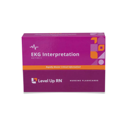
In this article, we cover the different sinus rhythms including normal sinus rhythm, sinus bradycardia, sinus tachycardia, as well as something called sinus arrhythmia. The EKG Interpretation video series follows along with our EKG Interpretation Flashcards, which are intended to help RN and PN nursing students study for nursing school exams, including the ATI, HESI, and NCLEX.
EKG Interpretation - Nursing Flashcards
What is a sinus rhythm?
The sinus node is one of the natural pacemakers of the heart, creating an electrical pulse that travels through the muscles in the heart, causing it to beat. A sinus rhythm is any cardiac rhythm where depolarization of the heart muscle begins at the sinus node.

Normal sinus rhythm
A normal sinus rhythm is considered the normal rhythm of a healthy heart, meaning the electrical pulse from the sinus node is exactly as expected. A normal sinus rhythm is the one you'd prefer to see on every patient!
A normal sinus rhythm on an EKG will show an equal distance from R wave to R wave and P wave to P wave. If you recall from our last article, the key characteristics of a normal sinus rhythm includes P waves that are upright and uniform in appearance, and there always being one P wave for each QRS complex.
Calculating heart rate for a normal sinus rhythm
Calculating the heart rate of a normal sinus rhythm is done by using the small box method. Using calipers, count the number of small boxes between R waves, then take 1500 and divide that by the number of small boxes. This will give you the number of beats per minute (BPM).
For example: if we count 25 small boxes between R waves, we would take 1500 divided by 25 to get 60 BPM. A normal heart rate should be between 60-100 BPM.

Sinus bradycardia
Sinus bradycardia happens when the sinus node does not send enough electrical impulses to the heart, resulting in a heart rate that is lower than 60 BPM. In general, the term bradycardia means a heart rate below 60 BPM. For some people, a heart rate below 60 BPM is completely normal, particularly in younger adults and athletes. However, for some it can be a sign that their heart is not circulating enough oxygenated blood throughout their body.
An EKG strip with sinus bradycardia would show a regular heart rhythm, with consistent distances between both R waves and P waves. P waves will be upright with a smooth and consistent appearance. PR intervals will be of normal duration, the QRS complexes will be narrow, and there will be only one P wave for each QRS complex.
Interventions
In terms of interventions for sinus bradycardia, if the patient is asymptomatic, then often interventions are not required. However, if the patient is symptomatic, then we can treat the patient with medications such as atropine or with an artificial pacemaker.
Calculating heart rate for sinus bradycardia
Sinus bradycardia will have a heart rhythm that is regular, but less frequent. We can use the small box method to calculate the heart rate. For example, if we count 45 small boxes between R waves, we take 1500 divided by 45 to get 33 BPM. Since the BPM is below 60, and the rhythm is regular, the patient has sinus bradycardia.

Sinus tachycardia
Sinus tachycardia is the opposite of sinus bradycardia and occurs when the sinus node sends too many electrical impulses in a certain amount of time, leading to a faster heart rate. While the electric pulse that’s causing the heart to beat may be normal, the pace of these beats is faster than usual. In general, a heart rate of over 100 BPM is considered tachycardia. Sinus tachycardia can increase the risk of serious complications, including heart failure, stroke, or cardiac arrest.
An EKG strip with sinus tachycardia will show a regular heart rhythm with consistent distances between R waves and P waves. P waves will be upright, the PR interval will be normal and the QRS complex will be narrow. There will also be a QRS complex for every P wave. Much like bradycardia, the pattern of the waves look normal, but the heart rate is what differs from the expected range.
Interventions
When a patient has sinus tachycardia, it is usually due to some underlying cause that needs to be diagnosed. The patient could have something like a fever, hypotension, pain, caffeine consumption, anxiety or other underlying issues. Interventions are focused on determining the underlying cause and addressing it.
Calculating heart rate for sinus tachycardia
Sinus tachycardia will have a heart rhythm that is regular, but more frequent. We can use the small box method to calculate the heart rate.
For example, if we count 13 small boxes between R waves, we take 1500 divided by 13 to get 115 BPM. Since the BPM is above 100, and the rhythm is regular, the patient has sinus tachycardia.

Sinus arrhythmia
The term Arrhythmia, in general, means a problem with the rate OR the rhythm of the heart. In our bradycardia section of this article, we describe a sinus rhythm with a regular rhythm and an irregular rate (too slow). In the tachycardia section of this article, we described a sinus rhythm with a regular rhythm and an irregular rate (too fast). But it's also possible to have a sinus rhythm with an irregular rhythm and either regular or irregular rate. That's the type of sinus arrhythmia that we will describe here.
Respiratory sinus arrhythmia, is a specific type of sinus arrhythmia in which the heart rate changes as you inhale and exhale. In other words, when you breathe in, your heart rate increases and when you exhale, it falls. This condition is not a cause for concern and is a naturally occurring heartbeat variation that is very common in young healthy adults and children.
An EKG strip that shows a sinus arrhythmia will have varying distances between R waves, while the other components on the EKG waveform will be as expected. The P waves are upright and smooth in appearance, the PR interval is normal, QRS complexes are narrow and there is a QRS complex for each P wave. The difference will be in the varying distances between R waves.
Calculating heart rate for sinus arrhythmia
When you have a sinus arrhythmia where the heart rhythm is irregular, you can't use the small box method because the rate rate is not the same across the length of the EKG strip. In this scenario, you need to use the 6-second strip method to determine the heart rate. The first step in calculating heart rate with the six-second strip method is to first ensure you are dealing with a 6-second EKG strip.
A 6-second strip is made up of 30 big boxes. Each big block is 0.2 seconds in duration, so 5 big blocks is equal 1 second in total duration (.2 x 5 = 1), meaning you would need a total of 30 big boxes to make a 6-second strip. Once you know you are dealing with a 6 second strip, you count the number of QRS complexes within those 6 seconds and multiply by 10.
For example, if we count 5 QRS complexes within a 6 second strip, we would get approximately 50 BPM.


4 comments
thank you educational
Being a former teacher/tutor ( Spanish, A&P, Micro , ESL) I feel you do an amazing job here. I was sort of lost in my ECG Tech class as I’m working full time night shift as PCA/STNA and thanks to you I GET IT!!!
Being a former teacher/tutor ( Spanish, A&P, Micro , ESL) I feel you do an amazing job here. I was sort of lost in my ECG Tech class as I’m working full time night shift as PCA/STNA and thanks to you I GET IT!!!
It would be worth mentioning that a resting HR below 60 is typical in athletes and some even go below 30 at night.