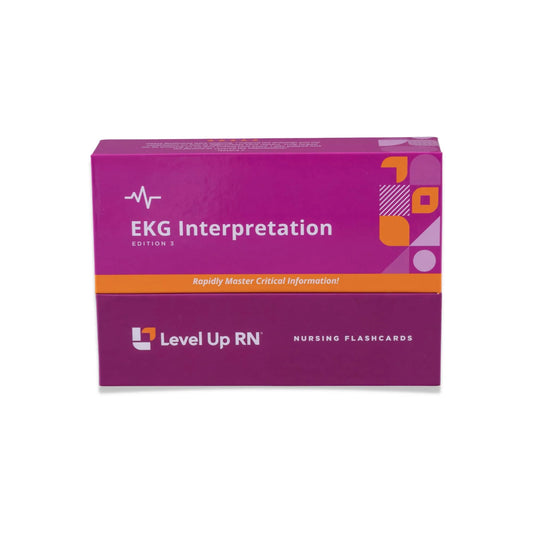
In this article, we cover EKG electrode placement when conducting a 12-lead EKG, the steps in the heart conduction system and some EKG basics to know. These basics are important to learn before you really dive into EKG interpretation. The EKG interpretation video series follows along with our EKG interpretation flashcards, which are intended to help RN and PN nursing students study for nursing school exams, including the ATI, HESI, and NCLEX.
EKG Interpretation - Nursing Flashcards
12-lead EKG placement
When performing a 12-lead EKG on a patient, we place 10 electrodes on their body. Four electrodes are placed on the patient's limbs and 6 on the patient's chest.
You might be wondering why there are 10 electrodes in a 12-lead EKG, and not 12! Leads actually don’t refer to the electrodes, but rather the perspectives through which we view the heart. So a "12-lead" means looking at the heart from 12 angles, which we obtain through 10 electrodes. There are some different configurations, but most 12-leads are done with 10 electrodes.
Lead placement on patient's limbs
- Lead LA = Left arm
- Lead LL = Left leg
- Lead RA = Right arm
- Lead RL = Right leg
Lead placement on patient’s chest
Leads placed on the patient's chest are placed relative to intercostal spaces (spaces between the ribs), and along vertical anatomical lines. You can think of the intercostal spaces like Y coordinates, or latitudes, and the vertical anatomical lines like X coordinates, or longitudes.
Lead V1
Place lead V1 on the 4th intercostal space (space below the 4th rib), at the right sternal border (where the sternum meets the ribs on the right side). This is the only lead that is placed on the right side of the chest.
Lead V2
Place lead V2 on the 4th intercostal space, at the left sternal border (where the sternum meets the ribs on the left side).
Lead V4
V4 comes before V3! This is simply because V3 is placed in between V2 and V4, so you need to place V4 first to know where V3 should go.
Place lead V4 on the 5th intercostal space (space below the 5th rib), at the midclavicular line (the line running vertically down the body from the midpoint of the clavicle). You may remember that this is the location where you can measure apical pulse.
Lead V3
Place lead V3 between V2 and V4.
Lead V5
Place lead V5 level with V4 at the anterior line (the line running vertically down the body where the armpit meets the chest in the front).
Lead V6
Place lead V6 level with V5 at the left mid-axillary line (the line running vertically down the body from the middle of the armpit).
Patient consideration
The chest EKG placement locations are often near a patient's inframammary fold, which is the point where the breast meets the chest. This area may be obscured by larger breasts that descend over that fold.
One important thing to keep in mind when performing an EKG on a patient with larger breasts is that you may need to lift the breast in order to place the leads correctly. We recommend bringing a second RN or CNA to assist you. It’s very important to communicate the procedure with your patient beforehand and explain that their breast will need to be lifted in order to place the electrodes.
Heart conduction system
Definitions of terms in the heart conduction system
| Term | Definition |
|---|---|
| Atria | Upper two chambers of the heart |
| Atrioventricular (AV) node | Electrical connection between the lower and upper chambers of the heart |
| Bundle branches (left, right) | Offshoots of the bundle of His leading to the left or right ventricle |
| Bundle of His | A bundle of muscle fibers that transmit electrical impulse |
| Depolarization | Contraction |
| Repolarization | Relaxation |
| Sinoatrial (SA) node or Sinus node | A group of cells in the heart that act as the heart's natural pacemaker by spontaneously producing an electrical impulse. |
| Ventricles | The lower two chambers of the heart |
| Ventricular myocardium | The muscle layer of the ventricles |
Steps in the heart conduction system
- The SA node initiates an electrical impulse, which stimulates the atria to depolarize. Atrial depolarization is recorded as a P wave on the EKG
- The electrical impulse travels down to the AV node, where there is a DELAY to allow the blood in the atria to empty into the ventricles. This pause is recorded as the PR segment on the EKG, shown as the short flat line after the P wave.
- The electrical impulse then travels down rapidly to the bundle of His, which transmits the impulse to the left and right bundle branches, where it travels to the ventricular myocardium. Depolarization of the ventricular myocardium triggers ventricular contraction. Ventricular depolarization is recorded as the QRS complex on the EKG. Atrial repolarization also occurs during this time.
- Repolarization of the ventricular myocardium then occurs. The ST segment represents the initial phase of ventricular repolarization, and the T wave represents the rapid phase of ventricular repolarization.
EKG basics
Box size on an EKG strip
EKG strips are split into big boxes, which are then each further split into smaller boxes.
Big box
Each big box on an EKG strip represents 0.2 seconds in duration.
- 5 big boxes = 1 second
- 30 big boxes = 6 seconds
- 300 big boxes = 60 seconds
Small box
Each small box on an EKG strip represents 0.04 seconds in duration.
- 5 small boxes = 1 big box (5 x 0.04 seconds = 0.2 seconds)
- 25 small boxes = 1 second
- 150 small boxes = 6 seconds
- 1500 boxes = 60 seconds
| Big boxes | Small boxes | Seconds |
|---|---|---|
| ⅕ | 1 | .04 or 1/25 |
| 1 | 5 | 0.2 or ⅕ |
| 5 | 25 | 1 |
| 30 | 150 | 6 |
| 300 | 1500 | 60 |
Normal duration and amplitude of EKG components
P wave
The amplitude of the P wave can be up to 2.5 mm (2.5 small boxes high) and duration between 0.06 - 0.12 seconds (1.5 - 3 small boxes).
PR interval
The duration of the PR interval is normally between 0.12 - 0.20 seconds (3-5 small boxes).
QRS Complex
The duration of a normal QRS complex is between 0.04 - 0.10 seconds. QRS complexes with a duration < 0.12 seconds (3 small boxes) are considered normal.
ST segment
The normal amplitude of the ST segment is usually isoelectric (0 mm), but may be between -0.5 and 1mm.
QT interval
The duration of a normal QT interval is between 0.36 and 0.44 seconds (9 - 11 small boxes).
T wave
The normal amplitude of the T wave is up to 10 mm (10 small boxes high).
| Component | Expected duration | Amplitude |
|---|---|---|
| P wave | 0.06 - 0.12 seconds | 2.5mm or 2.5 small boxes |
| PR Interval | 0.12 - 0.20 seconds | n/a |
| QRS complex | 0.04 - 0.10 seconds | n/a |
| ST segment | n/a | -0.5 - 1.0mm, or flat |
| QT interval | 0.36 - 0.44 seconds | n/a |
| T wave | n/a | 10mm, or 10 small boxes |


3 comments
This helps to review what my professor teaches.
Thanks very helpful lecture about ekg
Thank you very helpful.